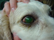
 Large corneal ulcer in a dog
|
cat breeds, feline breeds, Corneal Ulcer |
|
|
A corneal ulcer is an inflammatory condition of the cornea involving loss of its outer layer. It is very common in dogs and cats. It is also known as ulcerative keratitis.
Large corneal ulcer in a dog
The cornea is a transparent structure at the front of the eye. It refracts light and protects the contents of the eye. The cornea is about one-half to one millimeter thick in the dog and cat. The trigeminal nerve supplies the cornea via the long ciliary nerves. There are pain receptors in the outer layers and pressure receptors deeper.
Transparency is achieved through a lack of blood vessels, pigmentation, and keratin, and through the organization of the collagen fibers. The collagen fibers cross the full diameter of the cornea in a strictly parallel fashion and allow 99 percent of the light to pass through without scattering.
There are four important layers in the dog and cat cornea. The outer layer is the epithelium, which is 25 to 40 micrometers and five to seven cell layers thick. The epithelium holds the tear film in place and also prevents water from invading the cornea and disrupting the collagen fibers. This prevents corneal edema, which gives it a cloudy appearance. The epithelium sticks to the basement membrane, which also separates the epithelium from the stroma. The corneal stroma comprises 90 percent of the thickness of the cornea. It contains the collagen fibers organized into lamellae. The lamellae are in sheets which separate easily. Posterior to the stroma is Descemet's membrane, which is a basement membrane for the corneal endothelium. The endothelium is a single cell layer that separates the cornea from the aqueous humor.
An ulcer of the cornea heals by two methods: migration of surrounding epithelial cells followed by mitosis (dividing) of the cells, and introduction of blood vessels from the conjunctiva. Simple, small ulcers heal by the first method. However, larger or deeper ulcers often require the presence of blood vessels to supply inflammatory cells. White blood cells and fibroblasts produce granulation tissue and then scar tissue, effectively healing the cornea.
Corneal ulcers are one of the most common eye diseases in dogs. They are caused by trauma, detergent burns, and infections. Other eye conditions can cause corneal ulcers, such as entropion, distichia, corneal dystrophy, and keratoconjunctivitis sicca. Superficial ulcers involve a loss of part of the epithelium. Deep ulcers extend into or through the stroma and are can result in severe scarring and corneal perforation. Descemetoceles occur when the ulcer extends through the stroma. This type of ulcer is especially dangerous and can result in perforation. Corneal ulcers are painful due to nerve exposure, and can cause tearing, squinting, and pawing at the eye.
Diagnosis is through the use of fluorescein stain, which is taken up by exposed corneal stroma and appears green. With descemetoceles, Descemet's membrane will bulge forward and after staining will appear as a dark circle with a green boundary, because it doen not absorb the stain.
Treatment of corneal ulcers includes topical antibiotic therapy to prevent infection and pain medications, including topical atropine to stop spasms of the ciliary muscle. Superficial ulcers usually heal in less than a week. Deep ulcers and descemetoceles may require corneal suturing, conjunctival grafts or conjunctival flaps, soft contact lenses, or corneal transplant. Topical corticosteroids should never be used on any type of corneal ulcer because they prevent healing and will often make them worse.
Refractory corneal ulcers are superficial ulcers that heal poorly and tend to recur. They are also known as indolent ulcers or Boxer ulcers. They are caused by a defect in the basement membrane. They are recognized by undermined epithelium that surrounds the ulcer and easily peels back. Refractory corneal ulcers are most commonly seen in middle aged or older dogs and often occur in the other eye later. They are similar to Cogan's cystic dystrophy in humans.
Refractory corneal ulcers can take a long time to heal, sometimes months. Topical antibiotics are used continually to prevent infection. Pain medications are given as needed. Loose epithelium is removed with a dry cotton swab under topical anesthesia. This is in order to allow production of normal basement membrane and division of normal epithelium. Often further treatment is necessary, such as a keratotomy, which is superficial cutting or piercing of the cornea. There are two main types used in dogs: multiple punctate keratotomy (MPK) and grid keratotomy (GK). MPK involves making small superficial punctures into the cornea with a needle. GK is more commonly used and involves making parallel and perpindicular scratches in the corneal surface. Usually only topical anesthesia is necessary. By scoring the corneal surface, anchoring points are provided for attachment of new epithelium. Of course, these procedures should only be performed by a veterinarian, particularly one with some experience in this treatment.
Melting ulcers are a type of corneal ulcer involving progressive loss of stroma in a dissolving fashion. This is most commonly seen in Pseudomonas infection, but it can be caused by other types of bacteria or fungi. These infectious agents produce proteases and collagenases which break down the corneal stroma. Treatment includes antibiotics and collagenase inhibitors such as acetylcysteine and blood serum. Surgery may be necessary.
Corneal ulcers in cats can be caused by trauma, detergent burns, infections, and other eye diseases. One common cause not seen in dogs is infection with feline herpesvirus-1 (FHV-1). FHV-1 causes ulceration by direct infection of the epithelial cells. Lesions appear as dendritic (branching) ulcers. FHV-1 also suppresses healing of the cornea. Symptoms include conjunctivitis, squinting, eye discharge, and blood vessels on the cornea. It can cause severe scarring. Treatment is with topical antiviral drugs.